Horseshoe Crabs and Endotoxins
With over 4 billion people to date (March 2022) having received a Covid-19 vaccination, the human race owes a huge debt to countless researchers, healthcare workers and volunteers. Many of us would not be aware, however, that we also owe a debt of gratitude to a 450-million-year-old living fossil!
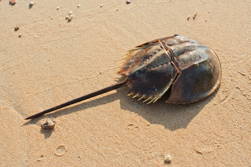
Meet the weird looking Horseshoe crab – a creature that ‘arrived on the scene’ 100 million years before the dinosaurs, isn’t actually a crab and has blue blood! Here’s a great video of them from the BBC.

This creature (Limulus polyphemus), not content with withstanding whatever the planet has thrown at it for multiple millennia, has helped saved lives and continues to do so. How? – the answer, remarkably, is in that blue blood!
What makes limulus blood so special?
Horseshoes crab’s blood is blue due to the presence of hemocyanin – where copper is used to carry oxygen rather than iron. These crabs have a very open circulatory system and swim in a bacterial rich environment. As such they are very susceptible to bacterial invasion and unlike warm blooded creatures, they cannot raise their temperature to kill of an infection. Instead, they have evolved a very sensitive method of detecting and dealing with infection. Amoebocytes in the blood detect bacterial invasion and trigger rapid clotting of the surrounding area, enmeshing the bacteria and plugging any wound.
This was reported by Fred Bang in 1953 [1], where it was shown that gram-negative bacteria will cause the blood of the horseshoe crab to turn into a semi-solid mass. It was demonstrated that even dead bacteria can trigger the clotting and was later shown to be the caused by lipopolysaccharide (LPS) endotoxins, a type of pyrogen [2].
The mechanism of clotting is now well studied and is known to involve a serine proteinase cascade. The components of the cascade are released from amoebocytes after LPS is detected. The clotting cascade is initiated by LPS binding to Factor C and finishes with coagulin polymerisation- shown below [3].

The Factor C coagulation pathway
Endotoxins
Endotoxins are lipopolysaccharide components of the outer membrane of Gram-negative bacteria such as E. coli. They are shed by growing bacteria and released when they die, are water soluble and heat resistant. In humans, LPS stimulates inflammation but excessive exposure can cause septic shock and organ failure [4]. For this reason, detecting endotoxin contamination is extremely important for anything that enters the body through surgery, injection or therapy.
The LAL test
During the 1960’s,Frederik Bang and Jack Levin developed a test from Limulus polyphemus blood that detected the presence of endotoxin [5]. This test (LAL – Limulus amoebocyte lysate) used osmotically lysed Limulus amoebocytes, the solution of which would gel upon endotoxin or bacterial addition. This test was cheaper, more sensitive and quicker than the established method of using rabbits for endotoxin assays and was approved for use in 1977 by the FDA. Modern tests still utilise freeze dried horseshoe crab derived lysate. The endotoxin measurement is then using one of three different methodologies: the qualitative gel clot (like the original developed test) and two quantitative tests; one based on a colour change and the other on turbidity.
Impact of the LAL test
The LAL test is now the primary method of testing all parenteral drugs, vaccines, intravenous fluids and implanted medical devices that come in contact with blood or cerebrospinal fluid. An estimated 70 million tests are performed per year. Johns Hopkins Bloomberg School of Public Health has placed the LAL test among the “100 Most Important Contributions to Public Health.”
Working with endotoxins at Peak Proteins
For proteins used as therapeutics, endotoxin levels must be reduced to extremely low levels to avoid unwanted cellular and immune responses. Endotoxin contamination of proteins can be problematic for cell-based assays and animal use, and can even affect in vitro assays. At Peak Proteins, we are experienced in minimising endotoxin levels where required by our clients. We can employ a low endotoxin workflow utilising sodium hydroxide washed equipment and low endotoxin water, and use endotoxin removal columns where required. We can provide endotoxin levels of purified proteins in the standard EU/ml, measured by a LAL test of where required by our clients. This is usually very successful in reducing endotoxin levels to an acceptable level in our HEK cells, CHO cells and insect cell expression systems. Expression in E. coli understandable generates a high source of endotoxin which can be almost impossible to reduce to the required level, so one option is to switch to one of our insect or mammalian expression systems.

Written by Martin Jennings, who has been fascinated by these crabs since finding their moults on a beach during a conference.
References
[1] F. B. Bang, “The effect of a marine bacterium on Limulus and the formation of blood clots.,” Biological Bulletin, vol. 105, pp. 361–362, 1953.
[2] J. LEVIN and F. B. BANG, “THE ROLE OF ENDOTOXIN IN THE EXTRACELLULAR COAGULATION OF LIMULUS BLOOD.,” Bull Johns Hopkins Hosp, vol. 115, pp. 265–274, Sep. 1964, Accessed: Feb. 28, 2022. [Online]. Available here.
[3] T. J. Novitsky, “Limulus amebocyte lysate (LAL) detection of endotoxin in human blood,” Journal of Endotoxin Research, vol. 1, no. 4, pp. 253–263, Sep. 1994, https://journals.sagepub.com/doi/10.1177/096805199400100407
[4] R. Karima, S. Matsumoto, H. Higashi, and K. Matsushima, “The molecular pathogenesis of endotoxic shock and organ failure,” Molecular Medicine Today, vol. 5, no. 3. Elsevier Current Trends, pp. 123–132, Mar. 01, 1999. https://pubmed.ncbi.nlm.nih.gov/10203736/
[5] J. Levin, P. A. Tomasulo, and R. S. Oser, “Detection of endotoxin in human blood and demonstration of an inhibitor,” The Journal of Laboratory and Clinical Medicine, vol. 75, no. 6, pp. 903–911, Jun. 1970, https://www.translationalres.com/article/0022-2143(70)90189-7/fulltext


